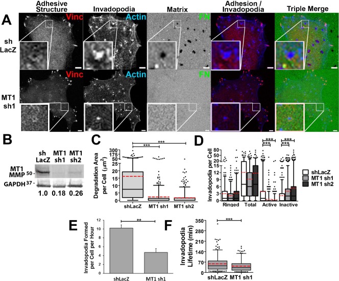
Fig. 6. MT1-MMP is required for invadopodium-associated ECM degradation, but not for adhesion ring localization.
(A–D) SCC61 cells stably expressing shRNA constructs against MT1-MMP (MT1 sh1 or sh2) or shLacZ control oligo were cultured on FITC-FN/gelatin substrates for 16 hours and then fixed and immunostained for vinculin (Vinc, red in merges) and actin (blue in merges). (A) Confocal images. Zoomed images show representative invadopodia for the condition. Scale bars = 5 µm. (B) Western blot of total cell lysates. Numbers indicate the ratio of MT1-MMP in each cell line normalized to the GAPDH loading control and then to shLacZ control. (C) Quantification of degradation area per cell. (D) Quantification of types of invadopodia per cell n≥60 cells per condition from ≥3 independent experiments. (E,F) Invadopodium formation (E) and lifetime (F) quantification of live-cell images of SCC61 cells stably expressing shLacZ or MT1 sh1 and transiently transfected with Tom-Tractin to mark invadopodia. n≥12 cells, ≥180 invadopodia per condition from ≥3 independent experiments. Error bars on invadopodium formation graphs indicate SEM. For C,D,F, box and whiskers show respectively the 25–75th and 5–95th percentiles with the dotted red line indicating the mean and the black line indicating the median. *P<0.05; **P<0.01, ***P<0.001.

