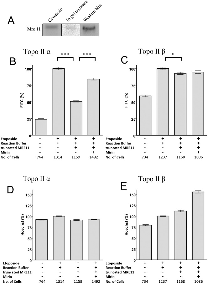
Fig. 3. Purified recombinant MRE11 removes topoisomerase IIα covalent complexes from genomic DNA.
(A). Coommassie, in-gel nuclease and Western analysis of recombinant MRE11. (B–E) K562 cells were treated with 100 µM etoposide for two hours. Slides bearing agarose-embedded cells (1–2×10⁶ cells in 100 µl 1% agarose in PBS spread across the slide surface) were incubated with MRE11 buffer or 1 µg MRE11 in MRE11 buffer in the presence or absence of the MRE11 nuclease inhibitor mirin and quantitative immunofluorescence was carried out for topoisomerase IIα or -β. (B,C) The mean FITC fluorescence for each nucleus was normalised to the 100 µM etoposide positive control, and the mean±SEM are shown. *** = p value, 0.0001, * = p value <0.05. (D,E) The mean hoechst fluorescence for each nucleus was normalised to the positive control, and the mean±SEM are shown.

