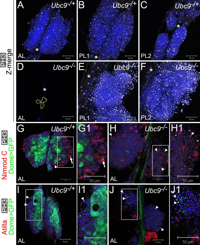
Fig. 4. Overproliferation of immature cells in Ubc9 lymph gland.
(A–F) Phosphorylated histone H3 (PH3, white) in 6.5–7-day L3 Ubc9−/+ AL, PL1, PL2 (A,B,C, respectively) and Ubc9−/− AL (D; remaining cells outlined), PL1, PL2 (E,F, respectively). Star marks DV. Optical Z-sections merged (A–C,E–F). Mutant PL1 and PL2 (E,F) are partially detached from the DV and misaligned; lobe orientation (top – anterior, bottom – posterior) is reverse of the DV. (G–J1) Dome>GFP (green), PH3 (white; arrowheads) and Nimrod C (red, G–H1), or Atilla (red, I–J1) in 6-day AL in Ubc9−/+ (G,G1,I,I1) and Ubc9−/− (H,H1,J,J1) animals. PH3/Nimrod C localization in the same cell (G1, arrow). Regions indicated in G,H,I,J magnified in G1,H1,I1,J1, respectively. Confocal sections (A–J1). Scale bars: 50 µm.

