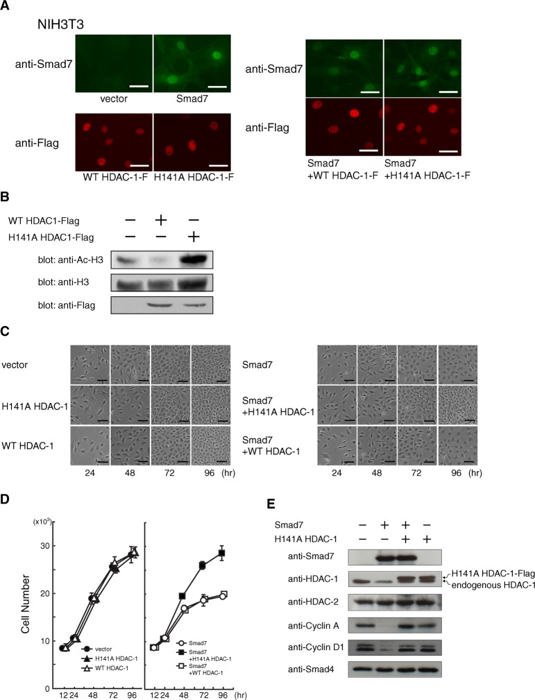
Fig. 3. Release of Smad7-induced cell cycle arrest by the H141A mutant of HDAC-1.
(A) Minimal effect on the level and localization of Smad7 when co-expressed with either wild-type or H141A HDAC-1. NIH 3T3 cells infected with combinations of retroviral vectors expressing the indicated protein: Smad7, wild-type HDAC-1, or an alanine substitution mutant for histidine 141 in HDAC-1 (H141A HDAC-1). Cells re-plated 48 h before fixation were single- or double-stained with rabbit α-Smad7 and mouse α-Flag antibody, followed by visualization with Alexa488-labeled α-rabbit Ig (green) and a Cy3-labeled α-mouse Ig (red) secondary antibody, respectively. Bars, 40 μm. (B) Effect of H141A HDAC-1 on the acetylation level of histone H3. NIH 3T3 cells were infected with control or retroviral vectors expressing Flag-tagged wild-type or H141A HDAC-1, and harvested 72 h later. Acetylation levels were examined by Western blotting using an antibody specific for acetylated histone H3 at both Lys9 and Lys13. Comparable protein levels were confirmed for total endogenous histone H3 in each sample and for exogenous wild-type and H141A HDAC-1 by using α-histone H3 and α-Flag, respectively. (C) Microscopic images of the cultures were taken to monitor the proliferation and morphology of NIH 3T3 cells expressing Smad7 and either wild-type or H141A HDAC-1. After 12 h of serial vector infection, cells were seeded (2×10⁵/a well of 6-well plate) and periodically photographed until 96 h under a phase-contrast microscope. Bars, 200 μm. (D) Effects of H141A HDAC-1 on Smad7-induced proliferation arrest. After 12 h of infection, NIH 3T3 cells (5×10³/a well of 96-well pate) were seeded to allow proliferation. The number of cells at 12, 24, 48, 72 and 96 h was measured as DNA content in each well. Transduced genes were as follows: •, two controls; ▴, H141A HDAC-1 and a control; △, wild-type HDAC-1 and a control; ◯, a control and Smad7; ▪, H141A HDAC-1 and Smad7; and □, wild-type HDAC-1 and Smad7. Data indicate means with S.D. from triplicate assays. (E) Effects of a dominant-negative HDAC-1 against Smad7 on the levels of various proteins. Cells infected with control, H141A HDAC-1, or Smad7 vectors were seeded (3×10⁵ cells/a 60-mm dish) and harvested at 72 h of culture. Western blots show the levels of exogenous Flag-tagged H141A HDAC-1 and Smad7, and of endogenous HDAC-1, HDAC-2, Cyclin A, Cyclin D1 and Smad4 proteins.

