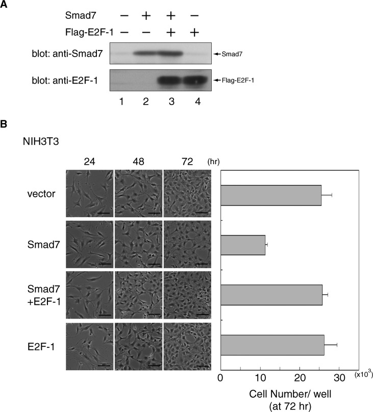
Fig. 6. Restoration of cell proliferation by E2F-1 from the Smad7-induced arrest in NIH 3T3 cells.
(A) Minimal effect of ectopic E2F-1 on the level of Smad7. NIH 3T3 cells were infected with retroviral vectors containing control, Smad7 or Flag-E2F-1, as indicated, and re-plated for further culture for another 72 h. The levels of Smad7 and Flag-E2F-1 were examined by Western blots of total cell extracts using α-E2F-1 and α-Smad7. (B) NIH 3T3 cells transduced in (A) were photographed periodically at the indicated time points to monitor their growth and morphology (see Fig. 3C legend). Bars, 200 μm. The number of cells in each culture after 72 h of re-plating was measured (see Fig. 3D legend). Data represent means with S.D. from triplicate experiments.

