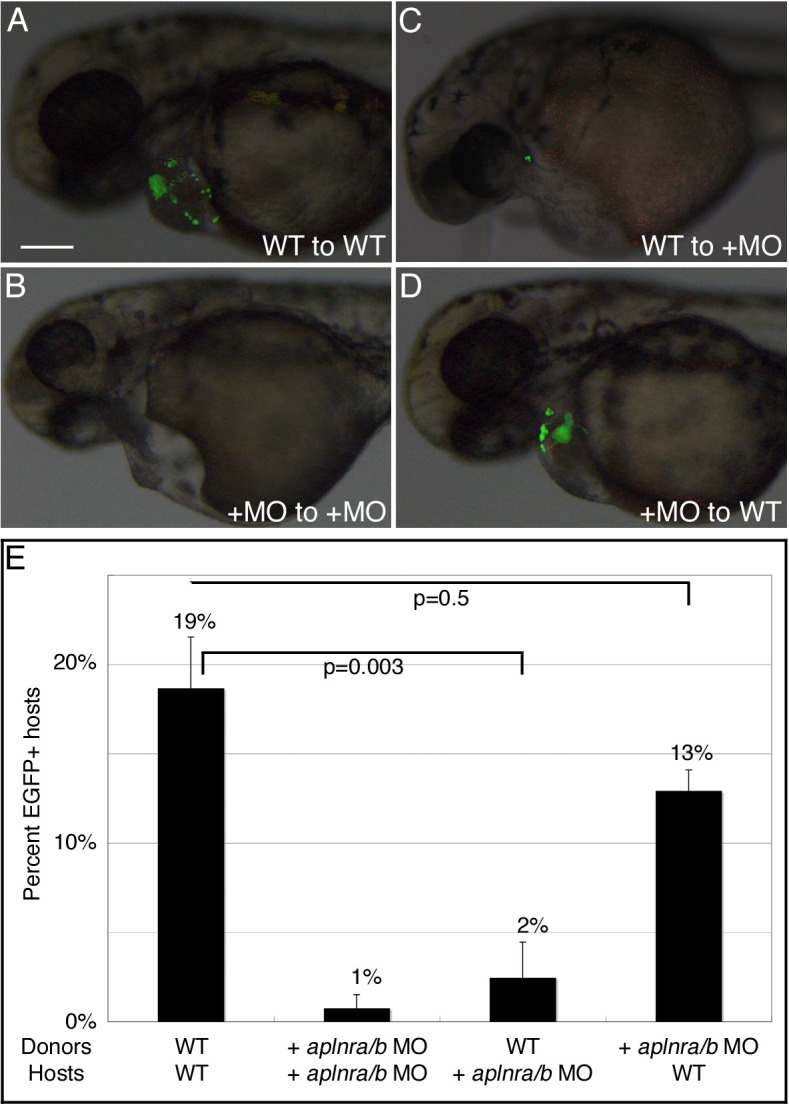
Fig. 3. Aplnr signaling functions cell-non-autonomously in the development of cardiomyocytes.

Transplantation of wildtype (WT) or aplnra/b MO-injected (+MO) myl7:EGFP donor embryos to the margin WT or aplnra/b MO-injected host embryos was carried out at 4hpf. (A–D) Representative host embryos at 2dpf following transplantation; lateral view, anterior to left. (C) wildtype cells are unable to contribute to the heart in aplnra/b morphant host embryos, whereas aplnra/b morphant cells are able to contribute to the heart in wildtype hosts (D). (E) Graph of percentage of host embryos with contribution from myl7:EGFP donor cells. N = 5, n = 236 for wildtype donors and hosts; N = 4, n = 130 for aplnra/b morphant donors and hosts; N = 4, n = 190 for wildtype donors and aplnra/b morphant hosts; N = 3, n = 190 for aplnra/b morphant donors and wildtype hosts. Views in (A–D) are lateral views, with anterior to the left. Scale bar represents 200μm (A).
