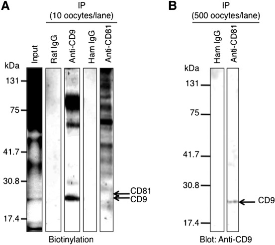
Fig. 4. Immunoprecipitation patterns of oocytes.

(A) Immunoprecipitation (IP) of oocyte lysates using anti-CD9 and anti-CD81. A total of 200 oocytes were collected from oviducts of superovulated female mice, and cumulus cells were removed from oocytes. Oocytes were biotinylated for 1 hour at 4°C and then lysed in 1% Brij 97-containing buffer for 3 hours at 4°C. This input lysate was next reacted with each anti-CD9 or anti-CD81 for 2 hours at 4°C, and precipitated with Sepharose beads conjugated with secondary Abs. After immunoprecipitation, the lysates corresponding to 10 oocytes were electrophoresed per lane. The preimmune rat IgG and hamster IgG (ham IgG) were concomitantly reacted with the oocyte lysates as negative controls. (B) Immunoblotting of the precipitate after reaction with anti-CD81. 500 oocytes/lane were collected from oviducts and lysed in Brij 97-containing buffer for 3 hours at 4°C. The lysates were reacted with anti-CD81 for 2 hours at 4°C and precipitated with Sepharose beads conjugated with secondary Ab. As a negative control, the oocyte lysates were precipitated with the preimmune hamster IgG (ham IgG). The precipitates corresponding to 500 oocytes were then electrophoresed per lane and immunoblotted with anti-CD9.
