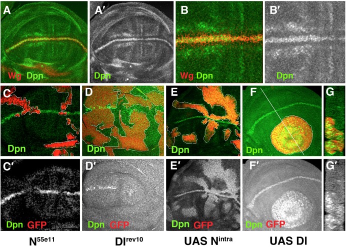
Fig. 1. Dpn expression is regulated by Notch signalling.

(A–G) Third instar larval wing discs stained with anti-Dpn (in green in A–G and grey in A′–G′) and anti-Wg (in red A,B and grey B′) antibodies. (A–B′) Dpn is expressed at high levels at the d/v boundary in the cells that express Wg. (B,B′) High magnification of panel A. (C,C′) Dpn is not expressed in N55e11 mutant clones (positively marked with GFP in red). (D,D′) In M+ Dlrev10 mutant clones (marked by the absence of GFP in red), Dpn is not expressed, except in some mutant cells at the border of the clone that were non-autonomously rescued by the adjacent wild type cells. (E,E′) Dpn is ectopically expressed in Nintra-expressing cells positively marked with GFP in red. (F–G′) In clones of Delta-expressing cells (marked by the expression of GFP in red), Dpn is expressed at high levels. (G,G′) A longitudinal cross-section at the position of the white line of panel F is shown. Mutant clones are outlined in white.
