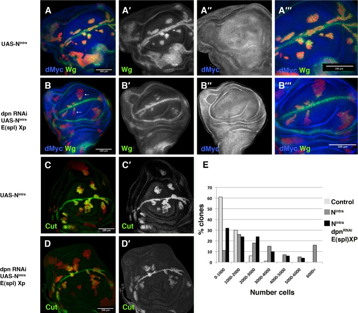
Fig. 6. E(spl)C and dpn mediate the functions of Notch signalling in regulating cell proliferation and the defining the d/v boundary.
(A–D′) Third instar larval wing discs stained with anti-Wg (in green in A,B,A‴,B‴, and grey in A′–B′), anti-Cut (green C,D, and grey C′,D′) and anti-dMyc (in blue A,B,A‴ ,B‴, and grey A″,B″) antibodies. Clones were positively marked with GFP in red (A,A‴,B,B‴,C,D). (A–A‴) In UAS-Nintra-expressing mutant cells, Wg was expressed at high levels throughout the wing blade. In these mutant cells, dMyc was down-regulated. (A‴) A high magnification view of panel A. (B–B‴) The up-regulation of Wg, caused by the ectopic activation of Nintra, was strongly suppressed in clones of UAS-dpnRNAi UAS-Nintra E(spl)Xp cells. In most of these clones, the ectopic expression of Wg was restricted to regions close to the wing margin (arrows), compared with the discs in A. (B‴) A high magnification view of panel B. (C,C′) The expression of Cut in discs contained clones of Nintra-expressing cells. (D,D′) In UAS-dpnRNAi UAS-Nintra E(spl)Xp mutant cells, Cut was ectopically expressed near the d/v boundary compared with C. (E) Quantitative analysis of the size of wild type (n = 59), UAS-Nintra (n = 79), and UAS-dpnRNAi UAS-Nintra E(spl)Xp (n = 50) mutant clones. Clone sizes were analysed using ImageJ (see Materials and Methods). The sizes of UAS-dpnRNAi and E(spl)Xp clones were similar to those of controls. Clones were induced at 60±12 h AEL and were analysed 96 h after induction.

