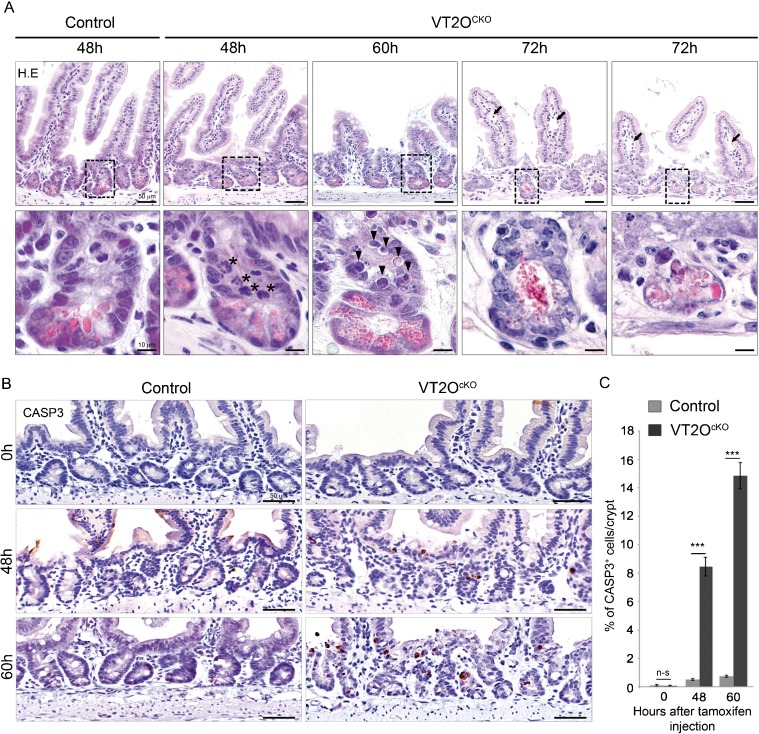
Fig. 3. Omcg1 loss of function in intestinal epithelium leads to massive apoptosis of crypt cells.
(A) Histological analysis of control and VT2OcKO mice intestine at different time points following TAM injection. Asterisks: abnormal mitotic cells; arrowheads: round acidophilic cell with apoptotic nuclear bodies, arrows: dilated lymph vessels showing oedematous mucosa. (B) Cleaved caspase-3 immunostaining 0, 48 and 60 hours after injection. (C) Mean (± SD) percentage of caspase-3 positive cells per crypt (n-s: non-significant; ***: p<0.001, Mann-Whitney-Wilcoxon test).

