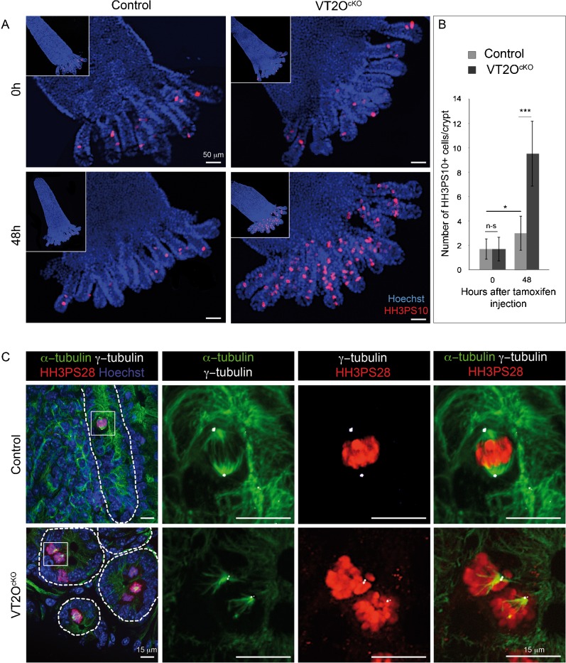
Fig. 5. Increased number of abnormal mitotic cells in Omcg1-deficient crypts.
(A) VT2OcKO and control villi with crypts attached at their bases were stained with Hoechst 33342 and anti-HH3PS10 antibody. (B) Number of mitotic cells per crypt was counted before and 48h after TAM injection (n-s: non-significant; *: 0.01 < p < 0.05, ***: p < 0.001, Mann-Whitney-Wilcoxon test). (C) Immunofluorescent staining on VT2OcKO and control intestine vibratome slices 48h after TAM injection. Hoechst 33342 (blue), anti-HH3PS28 antibody (red), anti-α-tubulin (green) and anti-γ-tubulin (white) were used for staining.

