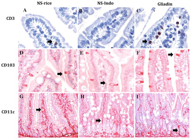
Fig. 2.
CD3, CD103, and CD11c immunostainings performed in small intestinal samples from HCD4/HLA-DQ8 transgenic mice. Representative pictures from tissue samples taken from non-sensitized mice orally challenged with rice cereal (NS-rice, n = 9) or indomethacin (NS-Indo, n = 5) as well as from animals sensitized and orally challenged with gliadin (Gliadin, n = 7). CD3 immunostaining show a brown reaction (a–c) while CD103 (d–f) and CD11c (g–i) immunostaining in red (a–f, 400×; and g–i, 200×). Specific brown or red staining is highlighted by black arrows

