Figure 1.
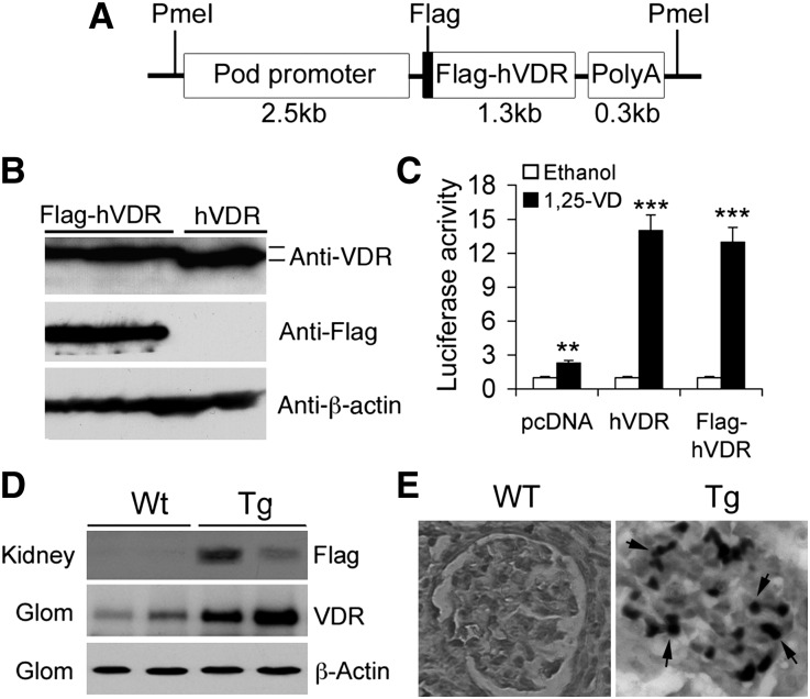
Generation of podocyte-specific hVDR transgenic mice. (A) Schematic illustration of podocin-Flag-hVDR-polyA DNA construct used for microinjection. (B) HEK293 cells were transfected with pcDNA-hVDR or pcDNA-Flag-hVDR and cell lysates were analyzed by Western blotting. Anti-VDR antibody recognizes both hVDR and Flag-hVDR, whereas anti-Flag antibody recognizes only Flag-hVDR, but not hVDR. (C) HEK293 cells cotransfected with p3xVDRE-Luc and pcDNA, pcDNA-hVDR, or pcDNA-Flag-hVDR are treated with ethanol or 1,25(OH)2D3. Luciferase reporter assays show that both hVDR and Flag-hVDR respond to 1,25(OH)2D3 stimulation and have the same activity. **P<0.01; ***P<0.001 versus corresponding ethanol, n=3. (D) Western blot analysis of total kidney lysates and purified glomerular (Glom) lysates from WT and Tg offspring, using anti-VDR antibodies or anti-Flag antibodies as indicated. (E) Immunostaining with anti-Flag antibodies shows Flag-positive (dark stain, arrows) podocytes in the glomerulus of Tg mice. Original magnification, ×400.

