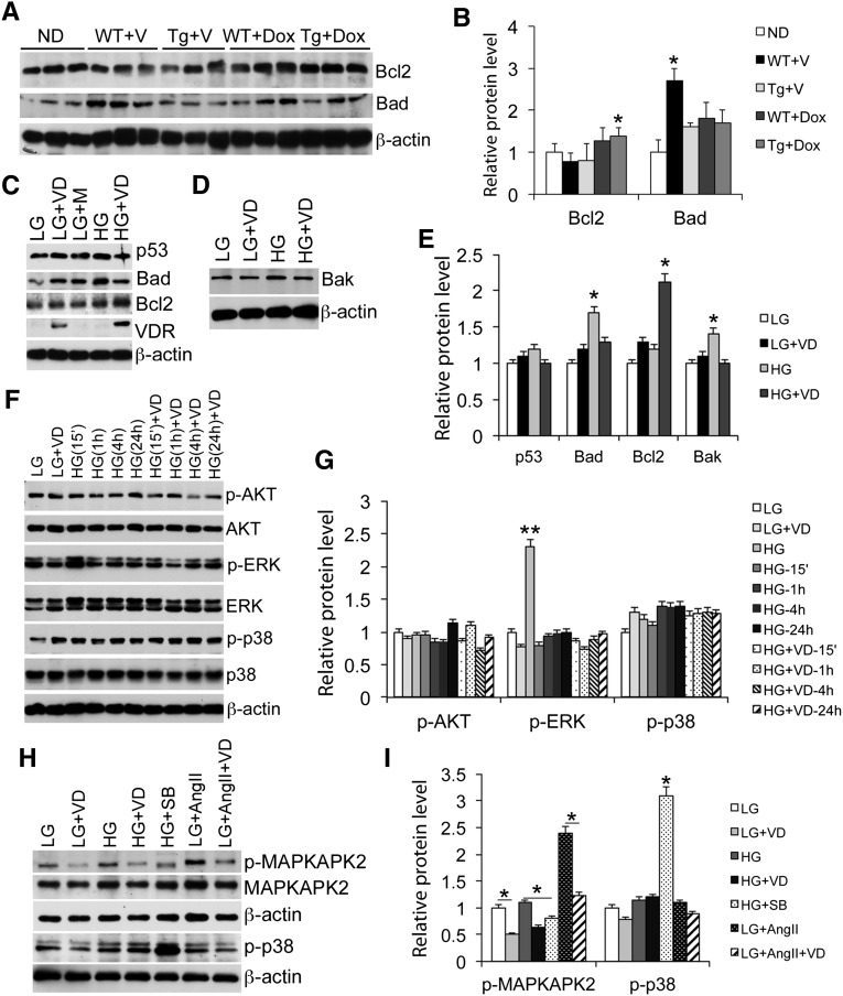
Figure 5.
Podocyte VDR signaling inhibits proapoptotic pathways. (A and B) Western blots (A) and densitometric quantitation (B) showing Bcl2 and Bad levels in glomerular lysates isolated from different mice. *P<0.05 versus the rest. (C–E) Western blots (C and D) and quantitation (E) showing levels of p53, Bad, Bcl2, and VDR in podocyte lysates under different cultural conditions as indicated. *P<0.05; **P<0.01 versus the rest. (F and G) Time course changes (F) and quantitation (G) in the levels of phospho-AKT, AKT, phospho-ERK, ERK, phospho-p38, and p38 in podocytes cultured in LG and HG conditions at different times (15 minutes to 24 hours) after 1,25-dihydroxyvitamin D treatment. **P<0.01 versus the rest. (H and I) Western blots (H) and quantitation (I) showing phosphorylation of MAPKAPK2 and p38 under different conditions. *P<0.05. VD, 1,25-dihydroxyvitamin D; M, mannitol; SB, SB203580. All Western blots are representative of at least three independent experiments.

