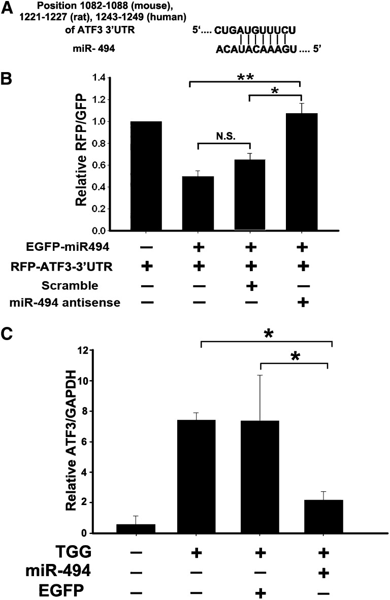
Figure 1.
Overexpression of miR-494 inhibits ATF3 expression in vitro. (A) Schematic representation of the putative miR-494 target sites in the 3′UTR of ATF3 of mouse, rat, or human. (B) ATF3 3′UTR activity assay. Fluorescent constructs containing EGFP–miR-494 and RFP-ATF3–3′UTR plasmids were cotransfected into 293T cells with or without scrambling or antisense plasmids. Fluorescent activity was determined 24 hours after transfection. The ratio of normalized sensor to control fluorescent activity is shown. The data are expressed as means ± SEM of three independent experiments. *P<0.05, **P<0.01. N.S., no significant difference. (C) Real-time PCR detected marked induction of miR-494, which dramatically decreased ATF3 levels in the NRK-52E cells induced by ATF3 inducer thapsigargin (TGG). The relative expression of the ATF3 was normalized to glyceraldehydes-3-phosphate dehydrogenase. *P<0.05 (n=3).

