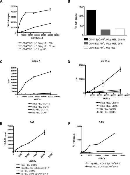
Figure 4. Presentation of blood-borne HEL by thymic DC and not TECs.
(A) Presentation of HEL by CD11c + DC isolated at 30 min and 36 h from B10.BR mice injected with 50μg HEL. (B) Corresponding presentation by 2-3×10⁴ TECs isolated from the same mice and cultured as in (A). For panels A-B, APCs were cultured for 24 h with 5×10⁴ 3A9 T-cell hybridoma and IL-2 production was measured by CTLL proliferation indicated by 3H-Thymidine incorporation. Proliferation of primary (C) 3A9α-/- and (D) LB11.3 T-cells in response to presentation by CD11c+ DC and CD45- TECs from mice injected with 50μg HEL. (E-F) Proliferation of primary 3A9 T-cells in response to presentation by (E) CD11c+ DC or (E, F). Sorted CD45-EpCAM+BP-1- mTECs isolated from mice injected with 1mg HEL. For primary proliferation, APCs were co-cultured with T-cells for 4 days and 3H-Thymidine incorporation was measured. In Panels C-F, mice were sacrificed 30 min post-injection and APCs isolated from un-injected B10.BR mice were used as controls.

