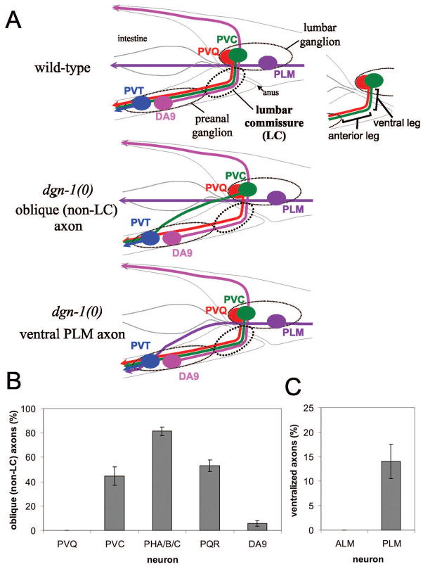
Figure 1. Defects in lumbar ganglion neural guidance in dgn-1 mutants.
(A) Several lumbar ganglion neurons, including PVQ (red) and PVC (green), project axons into the preanal ganglion through the lumbar commissure (LC), which has distinct ventral and anterior legs. The PLM (purple) axon in contrast projects anteriorly at a lateral position. The preanal ganglion neurons DA8 (not shown) and DA9 (pink) project axons dorsally through the left and right lumbar commissures, respectively; for convenience, DA9 is shown on the left in this schematic (upper panel). In dgn-1 null mutants, follower LC axons such as PVC sometimes fail to enter the lumbar commissure, instead taking an oblique route toward the preanal ganglion (middle panel), and PLM axons are occasionally directed ventrally toward the preanal ganglion (lower panel). (B) Oblique (non-LC) defects of specific lumbar commissure axons in dgn-1(cg121) null mutants is shown. For PHA/B/C, one or more of three axons per side could be defective (see Materials and Methods). (C) Ventralization of PLM axons in dgn-1(cg121). In B and C, percent of axons with guidance defects and the standard error of the proportion (error bars) is shown; N=100–232 axons scored for each neuron type.

