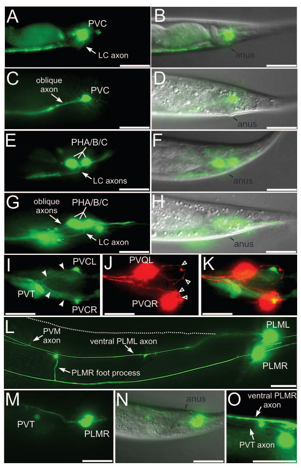
Figure 2. Axon guidance defects in lumbar neurons of dgn-1 mutants.
(A–D) Lateral views of lumbar commissure PVC (A) or PHA/B/C (E) axons in wild-type animals, and of oblique PVC (C) or PHA/B/C (G) axon trajectories in dgn-1(cg121) animals. One of the three PHA/B/C axons extends normally through the lumbar commissure in G. Overlays with the corresponding DIC images in (B,D,F,H) marking position of the anus. (I–K) Ventral view of a dgn-1(cg121) animal carrying markers for PVC (green, I), PVQ (red, J) and the preanal ganglion neuron PVT (green, I). Oblique PVC axons (I, filled arrowheads) extend directly toward the preanal ganglion, marked by PVT. The pioneer PVQ axons always traverse the lumbar commissure (J, open arrowheads) in dgn-1 mutants. K, overlay of I and J. In dgn-1(cg121), lumbar neuron cell bodies can be displaced anterior of their normal position, e.g. PVQL in J. (L) PLML axon in dgn-1(cg121) misrouted into the ventral nerve cord (marked by the entering PVM axon and the ventral cord foot process of PLMR). Normal lateral course of PLML is indicated (dotted line). In contrast the PLMR axon follows the normal lateral trajectory. (M–O) Subventral view of a dgn-1(cg121) animal carrying markers for PLM and PVT (green, M) and overlay with the DIC image (N), showing the misguided PLMR axon entering the ventral cord in the preanal ganglion region between PVT and the anus. In a different focal plane (O), the PVT and PLMR axons can be seen running parallel in the ventral cord. Bars: 20 μm.

