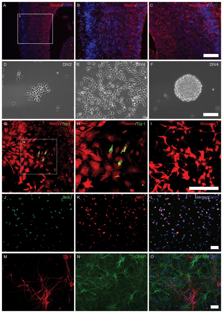
Figure 1. The isolation and identification of NSPCs from the fetal brain.
Images A–C: rat fetal brain sections were immunostained using anti-Nestin (red), anti-Musashi-1 (red), and DAPI (blue). Images D–F: in vitro cultured NSPCs; Images D–E: adherent monolayer NSPC cultures at days 2–4 in vitro; Image F: NSPC neurospheres in a non-coated culture dish. Images G–I: the expression of Nestin, Tuj-1, and Musashi-1 in cultured NSPCs (confocal microscopy); Images G–H: Nestin (red) and Tuj-1 (green); Image I: Musashi-1 (red). Images J–L: The proliferative potency of NSPCs; Image J: BrdU (green); Image K: Ki67 (red); Image L: a merged picture of image J, K, and DAPI (blue). Images M–O: Differentiation potency of NSPCs; Image M: Tuj-1 (red), a marker for newborn neurons; Image N: GFAP (green), a marker for astrocytes; Image O: a merged picture of Images M, N, and DAPI (blue). (Scale bars: 50 μm).

