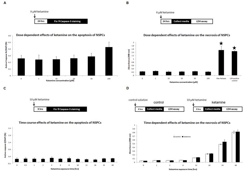
Figure 3. Effects of ketamine on apoptosis and necrosis in cultured NSPCs.
Panel A: dose-dependent responses of ketamine on apoptosis in cultured NSPCs. Ketamine exposure concentrations: X = 0, 1, 10, 20, 50, and 100 μM. Panel B: dose-dependent responses of ketamine (X = 0, 1, 10, 20, 50, or 100 μM) on necrosis in cultured NSPCs. Maximal release, cells were treated by Triton X-100; LDHpositive control, provided by the LDH assay kit. Absorbance values were measured at 490 nm. Stars represent significant differences compared to the control and all ketamine treatment groups. Panel C: time-dependent effects of ketamine on apoptosis of cultured NSPCs, as measured by percentage of active caspase-3+ cells in DAPI+ cells. Ketamine exposure duration: X = 0, 0.5, 1, 2, 4, 6, 8, 10, 12, 18, 24, and 48 hours. Panel D: time-dependent effects of ketamine on necrosis of cultured NSPCs using LDH assays. NSPC cultures were exposed to 10 μM of ketamine (black columns) for different durations (X = 0, 1, 2, 6, 12, 18, 24, and 48 hours) with parallel controls (white columns). No significant difference was found.

