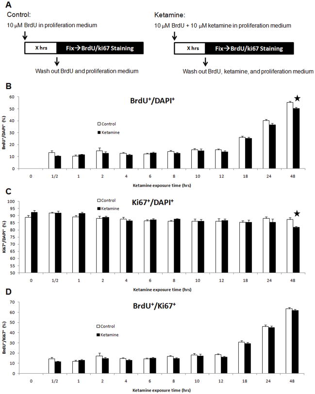
Figure 5. Time-dependent effects of ketamine on the proliferation of NSPCs.
Panel A: 10 μM of ketamine was added into cultures for different durations (X = 0,½, 1, 2, 4, 6, 8, 10, 12, 18, 24, and 48 hours) with parallel controls at each time point. Cultures were fixed and stained with anti-BrdU, anti-Ki67 antibodies and DAPI. Panels B–D: time-course response of ketamine on proliferation of cultured NSPCs. Panel B: the percentage of BrdU+ cells in total cells (DAPI+) vs. ketamine exposure time. Panel C: percentage of Ki67+ cells in the total cells (DAPI+) vs. ketamine exposure time. Panel D: the ratio of BrdU+ cells to Ki67+ cells vs. ketamine exposure time. (Stars represent the significant differences ketamine vs. control)

