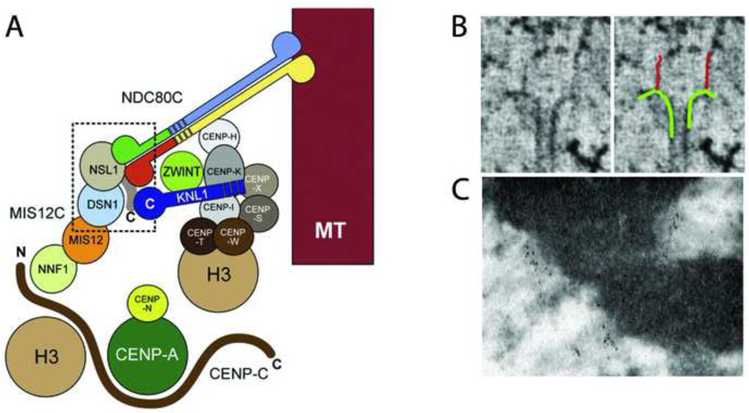
Figure 3. Molecular architecture of vertebrate kinetochores.
(A) Model of kinetochore protein interactions based upon structural and binding data. Adapted from [20] with permission. (B) Electron tomography images of long fibrils attaching microtubules to the inner kinetochore in PtK cells. Adapted from [18] with permission. (C) Immuno-EM localization of Ndc80 to the outer kinetochore. Three HeLa cell kinetochores are labeled with the 9G3 antibody to Ndc80 head. Note the linear concentrations of gold particles distal to the chromatin.

