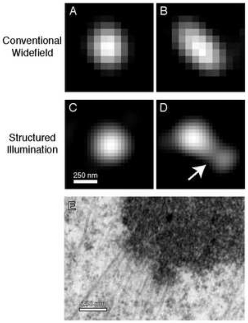
Figure 4. Details of kinetochore morphology revealed by super resolution microscopy.
(A) Labeled kinetochore proteins, such as the outer plate component Hec1, appear as round, diffraction limited point spread functions in conventional light microscopy. Methods based on centroid analysis fit the distribution of pixel intensities to a Gaussian function to find the center with sub-pixel accuracy. (B) Even in widefield, some Hec1 foci deviate from a PSF-like distribution of fluorescence, hinting at structural variation. (C and D) Hec1 immunostaining visualized with structured illumination microscopy (SIM). While some kinetochores still appear round, others exhibit a surprising amount of structural detail. In (D), a depression separates two apparent subdomains (arrow). (E) EM image of a kinetochore from a PtK1 cell highlighting the complexity of kinetochore morphology that can exist.

