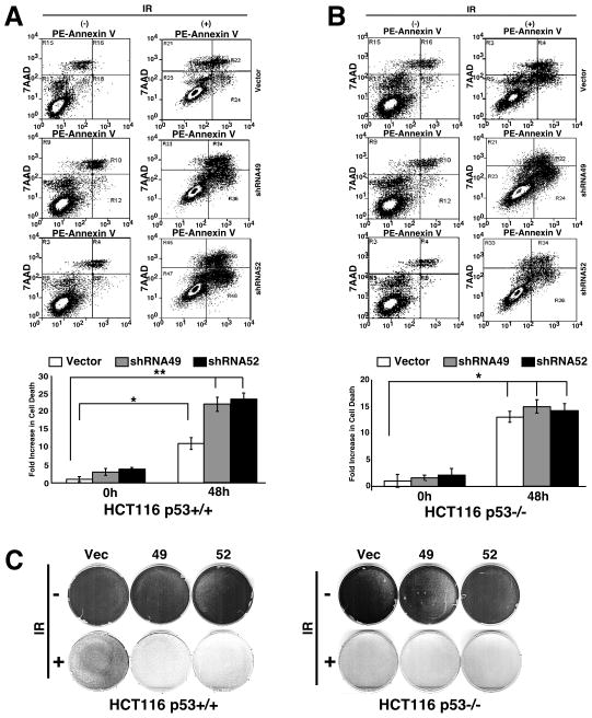
Figure 3. Knockdown of PTK6 in HCT116 cells leads to increased apoptosis and decreased survival following γ-irradiation.
HCT116 p53+/+ (A) and p53−/− (B) cells containing empty vector shRNA (Vector) or one of two different shRNAs (shRNA49, shRNA52) were stained with PE Annexin V and 7AAD and assayed by flow cytometry 0 (−) and 48 (+) hours post-20 Gy γ-irradiation. (A) Quantification of fold-increase in cell death for HCT116 p53+/+ (*P< 0.006, **P< 0.002; Bars +/− SD) and (B) HCT116 p53−/−(*P< 0.004; Bars +/− SD) shown in plots. (C) Colony formation assays were performed after seven days with HCT116 p53+/+ and p53−/− cells containing empty vector (Vector) or one of two different shRNAs (shRNA49, shRNA52) that target PTK6. Cells were untreated (− IR) or exposed to 20 Gy of γ-irradiation (+ IR).

