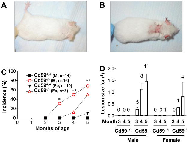
Figure 2.
CD59a−/− -MRL/lpr mice developed autoimmune dermatitis with high incidence and severity. Representative pictures of a CD59a+/+-MRL/lpr mouse at 5 month of age (A) showing the lack of skin disease and of an age-matched CD59a−/− -MRL/lpr mouse (B) showing severe skin disease. (C) Percentage of Cd59−/−-MRL/lpr (CD59a−/−) and Cd59+/+-MRL/lpr (CD59a+/+) mice with visible open skin lesions at 3, 4, and 5 months of age. Numbers of mice are indicated on the graph. * P<0.05, ** P<0.01, compared with male Cd59+/+-MRL/lpr mice. Fisher’s exact test. (D). Average size of open skin lesions (mean ± SED). Data are only for mice with open skin lesions (numbers indicated above the columns) among the same mice studied in C.

