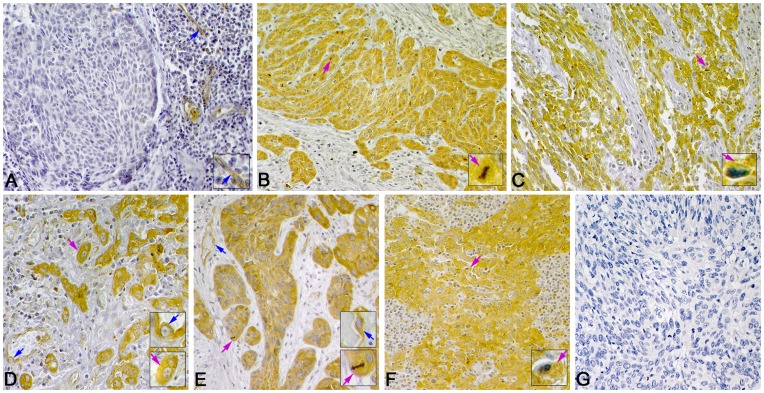
Figure 5. Representative immunohistochemistry staining of paxillin in LSCC.
Positive immunostaining of paxillin mainly in the cytoplasm of tumor cells (pink arrows) and in endothelial cells of microvessels (blue arrows). (A) Negative expression of paxiliin in well-differentiated LSCC. (B) High expression of paxillin in well-differentiated, (C) poorly differentiated, (D) supraglottic, (E) glottic, and (F) subglottic LSCC. (G) Negative control staining (PBS) of paxillin. (Magnification 400×, insets 1000×).

