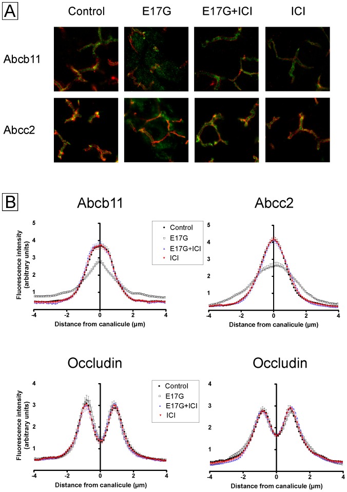
Figure 9. Estrogen Receptor inhibition prevents estradiol-17ß-d-glucuronide (E17G)-induced endocytic internalization of Abcb11 and Abcc2 in perfused rat liver.
Panel A: Confocal images of E17G-induced internalization of Abcb11 and Abcc2 and protection by ICI 182,780 (ICI). Representative confocal images of immunostained liver samples displaying a containing of Abcb11 (green) and occludin (red) (upper images), and Abcc2 (red) and occludin (green) (lower images). In control livers, both Abcb11 and Abcc2 were mainly confined to the canalicular space delineated by the tight junction-associated protein occludin. Following E17G (3 µmol/liver), some canaliculi show intracellular fluorescence associated with Abcb11 or Abcc2 at a greater distance from the canalicular membrane, consistent with their delocalization. ICI (0.5 µM, 15 min previous to E17G) prevented the internalization of canalicular transporters, as illustrated by a control-like pattern of Abcb11 and Abcc2 distribution. ICI itself did not induce any changes in transporters localization. Panel B: Densitometric analysis of fluorescence intensity profile of Abcb11, Abcc2 and occluding. Graphs represent the intensity of fluorescence associated with the transporters along an 8-µm line (from −4 µm to +4 µm of the canalicular center) perpendicular to the canaliculus. In control livers, transporter-associated fluorescence was concentrated in the canalicular space. E17G-induced internalization of transporters from the canalicular membrane (P<0.01 versus control) was detected as a decrease in the fluorescence intensity in the canalicular area together with an increased fluorescence at a greater distance from the canaliculus. Distribution profiles of livers treated with E17G+ICI was similar to control and indicated a significantly decreased of Abcb11 and Abcc2 internalization (P<0.01 versus E17G). (n = 20–50 canaliculi per preparation, three independent preparations). Statistical analysis of the distribution profiles of occluding, used to demarcate limits of the canaliculi, showed no changes in the normal distribution by any of the treatments.

