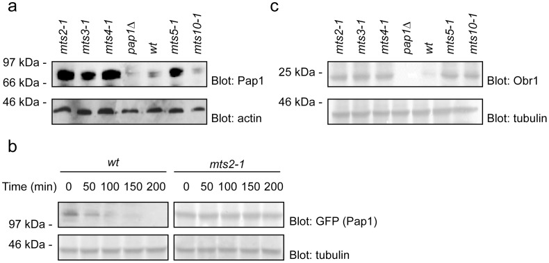
Figure 2. Stabilization of Pap1 in the mts mutants leads increased obr1+ expression.
(a) To compare the steady state levels of Pap1 cell extracts of the indicated strains were prepared and analyzed by SDS-PAGE and Western blotting using antibodies to Pap1. Actin served as a loading control. Compared to wild type cells, the Pap1 levels were increased in the proteasome mutants, but not in the mts10-1 (crm1) mutant. A pap1Δ mutant was included as a control. (b) The degradation kinetics of GFP-tagged Pap1 was followed by blotting of wild type (wt) and mts2-1 cultures treated with cycloheximide (CHX). α-tubulin served as a loading control. In wild type cells Pap1 was rapidly degraded with a half-life of about 50 minutes. In the mts2-1 background Pap1 was stabilized (c) To compare the steady state levels of the Pap1 target Obr1 cell extracts of the indicated strains were prepared and analyzed by SDS-PAGE and Western blotting using antibodies to Obr1. Tubulin served as a loading control. Compared to wild type cells, the Obr1 levels were increased in the proteasome mutants and, as expected, in the mts10-1 (crm1) mutant. No Obr1 was detected in the pap1Δ mutant.

