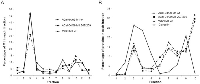
Figure 7. The pH1N1 M1 protein associates as efficiency with the plasma membrane and lipid raft domains as other influenza M1 proteins.
293 T cells were transfected with WT ACal-04/09 M1, ACal-04/09 M1 207/209 and WSN M1 protein expression plasmids. At 24 hpi, cells were (A) homogenized with a 27 gauge needle, mixed with 70% sucrose, and subject to membrane flotation analysis or (B) lysed in 0.5% Triton×100 and mixed with 70% sucrose for lipid raft isolation. Fractions were collected from the top of each gradient following ultracentrifugation, separated by gel electrophoresis and analyzed by Western Blot for M1 protein. Bands intensities were quantified using BioRad Software.

