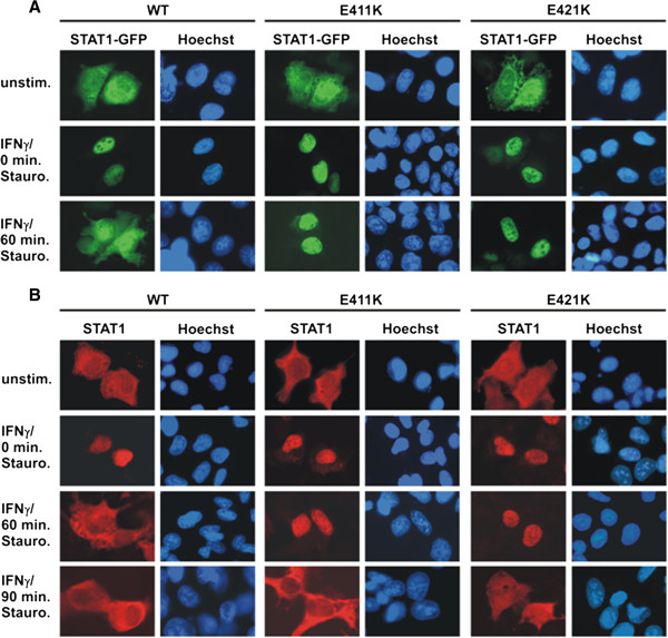
Figure 5.

STAT1 DNA-binding mutants with decreased dissociation from DNA exhibit a prolonged nuclear accumulation phase. (A) Localization of GFP-tagged STAT1 variants in HeLa cells expressing either wild-type or mutant STAT1, in which glutamic acid residues at positions 411 and 421 were substituted for lysine. The cells were left unstimulated (top panel), stimulated with IFNγ for 45 min (middle panel), or pre-treated with IFNγ and subsequently exposed to 500 nM staurosporine for an additional 60 min (bottom panel). Fluorescence micrographs demonstrate the intracellular STAT1-GFP distribution before and after cytokine stimulation, as well as the effects of staurosporine treatment. Corresponding Hoechst-stained nuclei are shown. Note that, in the presence of the kinase blocker staurosporine, both glutamyl mutants remained in the nuclear compartment, whereas in cells expressing wild-type STAT1 the same treatment resulted in the collapse of STAT1 nuclear accumulation. (B) Time course of the collapse of nuclear accumulation in U3A cells expressing the indicated untagged STAT1 variants. Cells were either left untreated or treated for 45 min with IFNγ followed by subsequently exposure to 500 nM staurosporine for 0, 60 and 90 min, respectively. The nuclei of the methanol-fixed cells were stained with Hoechst dye and the samples processed for immunocytochemical detection of STAT1 using a pan-STAT1 antibody and appropriate Cy3-labeled secondary antibody.
