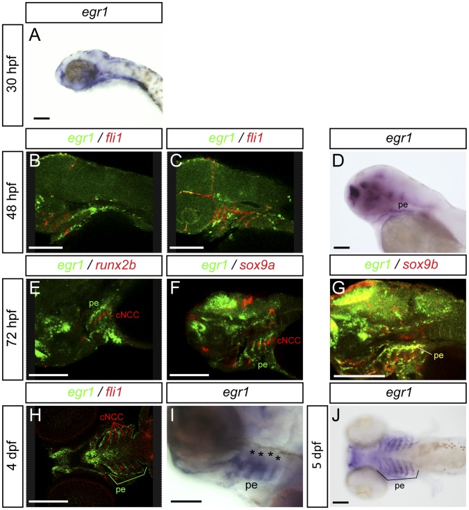
Figure 3. Expression of egr1 in the pharyngeal region between 30 hpf to 5 dpf is restricted to endoderm and epithelium.
Lateral (A–G,I) and ventral (H,J) views, anterior to the left. Scale bars 100 µm. Images of double in situ hybridizations were taken by confocal microscopy and pictures of individual Z-sections are shown. (A) egr1 transcripts are observed in the pharyngeal region starting at 30 hpf in endoderm. (B,C) At 48 hpf, double in situ hybridization for egr1 (green) and fli1 (red); egr1 transcripts are localized in pharyngeal endoderm and do not colocalize with fli1 mRNA in pharyngeal cartilage precursor cells. (D) egr1 is expressed in pharyngeal endoderm. (E–G) At 3 dpf, egr1 (green) does not colocalize with runx2b (red) (E) or sox9a (red) (F) in cartilage, while (G) egr1 (green) mRNAs colocalize with those for the pharyngeal endoderm marker sox9b (red). (H) At 4 dpf, egr1 (green) is never expressed in cells in pharyngeal cartilage precursor cells expressing fli1 (red). (I) Expression of egr1 at 4 dpf in pharyngeal endoderm. (J) At 5 dpf, egr1 is still expressed in pharyngeal endoderm (stars) and not in pharyngeal cartilage. Pharyngeal endoderm (pe), cranial neural crest cells (cNCC).

