
Figure 2. Reduction of Zfp521 enhances adipogenic potential in vivo.
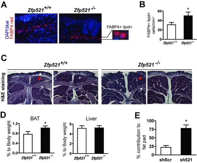
(A) Sections of the subcutaneous region of e18.5 Zfp521+/+ and Zfp521−/− embryos were stained with anti-FABP4 (red) and DAPI (blue). The magnified section shows representative FABP4+, lipid-filled cells. (B) ∼20–25 images per embryo were used to quantify the number of lipid-filled and FABP4-positive adipocytes. Zfp521+/+, n = 4 embryos; Zfp521−/−, n = 6 embryos; *p<0.05. (C, D) Representative histology (HE staining) of the interscapular BAT region from Zfp521+/+ and Zfp521−/− e18.5 embryos (red arrowhead, interscapular BAT; white arrow, skeletal muscle) (C) and BAT and liver weight relative to total body weight (D). Data presented as mean ± SD, Zfp521+/+ (n = 9); Zfp521−/− (n = 15), *p<0.05. (E) shZfp521 and scrambled hairpin (shScr) expressing F442A cells were mixed and injected into nude mice. Percentage contribution of shScr and sh521-expressing cells in fat pads determined using specific Q-PCR (see text for details). Data presented as mean ± SD, n = 11, *p<0.05.