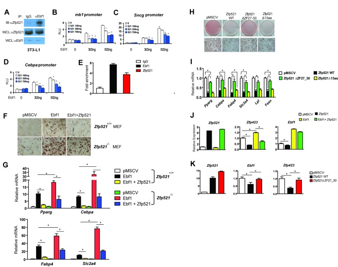
Figure 4. Zfp521 inhibits Ebf1 transcriptional activity through physical interaction.
(A) 3T3-L1 preadipocytes were harvested and endogenous Ebf1 was immunoprecipitated using anti-Ebf1 beads and blotted with normal goat-IgG or anti-Zfp521. (B–D) 3T3-L1 preadipocytes were co-transfected with vectors expressing Flag-Zfp521, Myc-Ebf1, and various reporter plasmids containing the mb1-promoter (B), Sncg-promoter (C), or Cebpa-promoter (D). At 24 h after transfection, luciferase activity was normalized to β-galactosidase activity. Data presented as mean ± SD, n = 4, *p<0.05. (E) 3T3-L1 preadipocytes were stably transduced with Flag-Ebf1 or Flag-Zfp521. ChIP assay was performed on 3T3-L1 cells that were treated with DMI for 1 h using anti-Flag antibody or an IgG control using PCR primers directed at regions of the Cebpa containing the putative Ebf sites. (F, G) Immortalized Zfp521+/+ and Zfp521−/− MEFs were transduced with a retrovirus expressing Ebf1, Zfp521, or empty vector and differentiated prior to staining with oil red-O after 8 d (F) and adipocyte gene expression was measured by Q-PCR (G). (H, I) C3H10T1/2 cells were transduced with a retrovirus expressing Zfp521WT, Zfp521ΔZF27-30, Zfp521Δ13aa, or empty pMSCV vector. Cells were differentiated with DMIR and stained with oil red-O and gene expression was measured on day 6. (J) Zfp521 and Ebf1 were expressed in C3H10T1/2 cells alone or in combination, and expression of Zfp521, Ebf1, and Zfp423 was determined by Q-PCR. (K) Zfp521, Zfp521Δ27-30, or pMSCV was expressed in C3H10T1/2 cells and expression of Zfp521, Ebf1, and Zfp423 was determined by Q-PCR. Data presented as mean ± SD, n = 3, *p<0.05.

