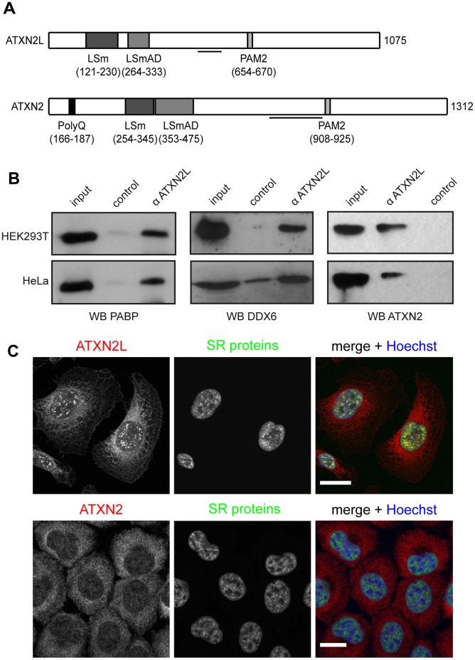
Figure 1. ATXN2L is found in association with PABP, DDX6 and ATXN2. A).
Scheme of the domain architecture of ATXN2L and ATXN2 highlighting conserved functional motifs. Lines below indicate antibody epitopes for anti-ATXN2L and anti-ATXN2 (BD Biosciences). B) Cell lysates were prepared from HEK293T and HeLa cells as described in Materials and Methods. Co-immunoprecipitation experiments were carried out with an anti-ATXN2L antibody, and precipitated proteins were detected using specific antibodies against PABP, DDX6 or ATXN2 (BD Biosciences). C) HeLa cells were fixed and ATXN2L or ATXN2 were stained with an anti-ATXN2L antibody or an anti-ATXN2 antibody (Sigma, red). SR-splicing proteins were stained using an anti-SR antibody (green). Nuclei were visualized by Hoechst staining (blue). Scale bars represent 20 µm.

