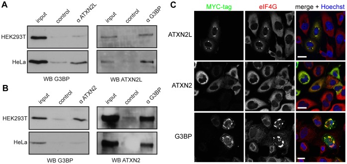
Figure 4. ATXN2L overexpression induces the formation of SGs. A).
For co-immunoprecipitation experiments cell lysates were prepared from HEK293T and HeLa cells as described in Materials and Methods, and experiments were carried out with either anti-ATXN2L or anti-G3BP antibody. Precipitated proteins were detected with anti-G3BP or anti-ATXN2L. B) Co-immunoprecipitation experiments were carried out with either anti-ATXN2 (Bethyl) or anti-G3BP antibody. Precipitated proteins were detected with anti-G3BP or anti-ATXN2 (Bethyl). C) HeLa cells were transfected with the expression plasmids RSV-ATXN2L-MYC, pCMV-MYC-ATXN2-Q22 or pCMV-MYC-G3BP1 and incubated for 48 hours to allow expression of the respective fusion proteins. Afterward, cells were fixed and stained for the MYC-tag (Millipore, green) and eIF4G (red) to monitor induction of SGs. Nuclei were stained using Hoechst (blue). Scale bars represent 20 µm.

