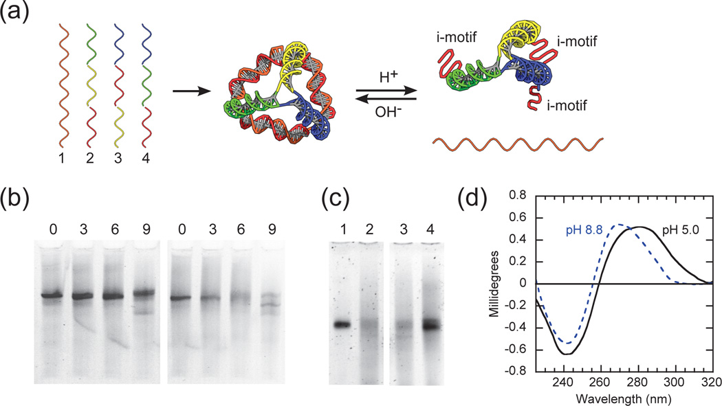
Fig. 1.
(a) Schematic of pH-triggered conformational changes of an i-motif DNA pyramid. The i-motif sequence is colored red and the complementary sequence is colored orange. Otherwise, identical colored regions are complementary to each other. (b) Mismatch-controlled conformational changes of i-motif DNA pyramids at different pH. The left and right gel are at alkaline (pH 8.8, unless otherwise stated) and acidic conditions (pH 5.0, unless otherwise stated), respectively. In both gels the lane numbers indicate the number of mismatches in strand 1. (c) pH-controlled assembly of i-motif DNA pyramids. Switching from alkaline to acidic conditions (lanes 1 and 2), and vice versa (lanes 3 and 4); (d) Circular dichroism (CD) of i-motif DNA pyramids at different pH.

