Fig. 3.
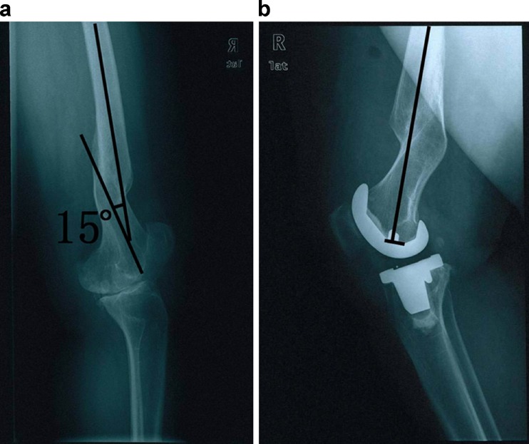
A 38-year-old woman who had had arthritis with extra-articular deformity. a Preoperative radiograph showing 15° sagittal deformity caused by malunited distal femur fracture. b Postoperative lateral radiograph made 2 years after one-stage TKA with intra-articular bone resection and soft tissue balancing, showing excellent alignment of the femoral component and no radiolucency about the component

