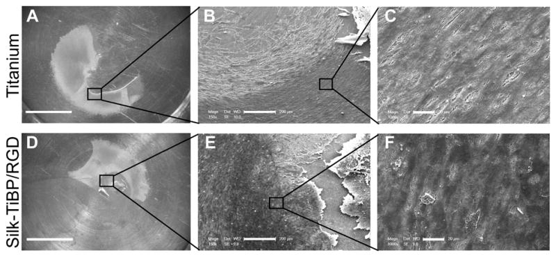
Figure 6. Endothelial tissue culture.

Organotypic tissue culture was realized on unmodified titanium discs (a, b, c) or functionalized with silk-TiBP/RGD 5.3% (d,e,f). Images a and d show the tissue expanding on titanium surface. SEM micrographs of explant at low magnification (b,e) and higher magnification (c,f) reveal the growing cells layer. Scale bars: a,d: 5mm; b,e: 200μm; c,f: 20μm.
