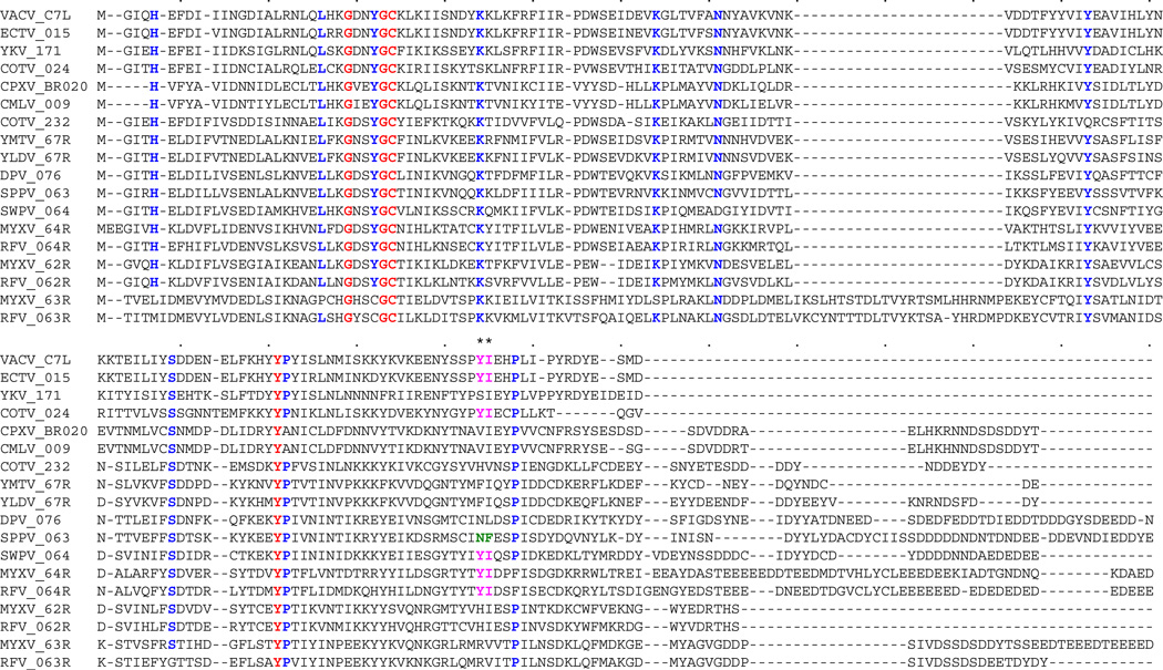
Figure 1.
Multiple sequence alignment of poxvirus C7L family members. A multiple sequence alignment of poxvirus C7L orthologs (see Table 1 for abbreviations) that show less than 93% amino acid sequence identity with one another by pairwise comparison was carried out using MAFT. Residues that are 100% conserved are shown in red, residues that are conserved in ≥90% of sequences are shown in blue. Residues 134 (N = Asparagine) and 135 (F = Phenylalanine) of sheeppox virus (SPPV) 063 are highlighted in green, and corresponding residues Y (Tyrosine) and I (Isoleucine) that were introduced into SPPV 063 and rescued virus replication in mouse 3T3 cells [19**] are shown in purple.

