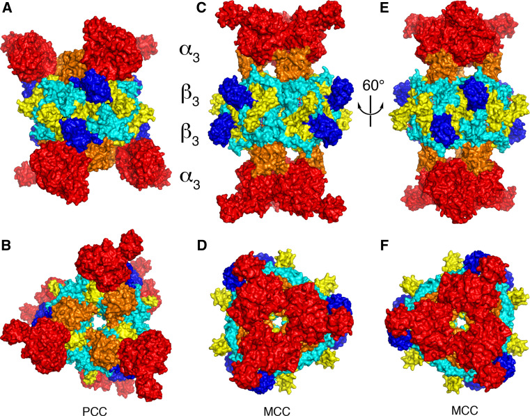
Fig. 6.
Striking differences in the overall architecture of the holoenzymes of PCC and MCC. a Crystal structure of the bacterial PCC holoenzyme [22], viewed down the twofold symmetry axis within a β2 dimer. The domains are colored as in Fig. 2. The four layers of the structure are indicated. b Structure of the PCC holoenzyme, viewed down the threefold symmetry axis. c Crystal structure of the P. aeruginosa MCC holoenzyme [23], viewed down the twofold axis within a β2 dimer. d Structure of the MCC holoenzyme, viewed down the threefold axis. e Structure of the MCC holoenzyme, after a ~60° rotation around the vertical axis from c. The view is down the twofold axis relating two β2 dimers. f Structure of the MCC holoenzyme, after a ~60° counterclockwise rotation from panel D

