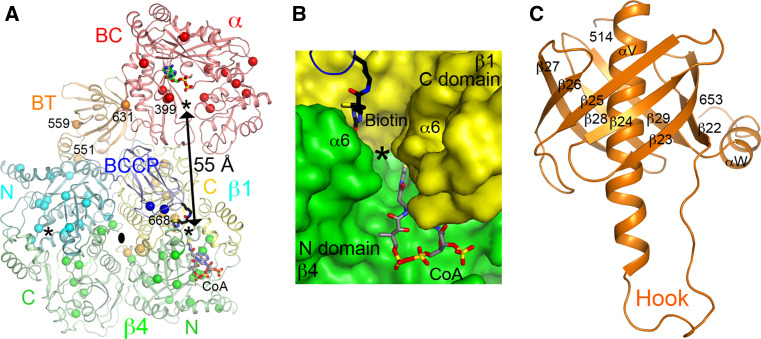
Fig. 7.
The active sites of PCC. a Relationship between the BC and CT active sites (indicated with the asterisks) in the PCC holoenzyme. The CT active site is located at the interface of a β2 dimer, with the β subunit from the bottom layer colored in green. Sites of disease-causing missense mutations are indicated with the spheres. The third asterisk indicates the other active site of the CT dimer. b Molecular surface of the active site region of CT. The observed position of biotin is shown (stick model in black). The position of CoA (gray) is modeled based on that in the structure of the yeast ACC CT domain [169]. c Structure of the BT domain of the bacterial PCC α subunit. The hook region is labeled

