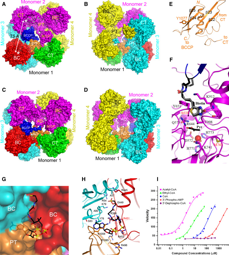
Fig. 9.
Structure of PC. a Crystal structure of SaPC tetramer [21]. The domains of monomer 1 are colored as in Fig. 2, and the other three monomers are in magenta, cyan and yellow. The BC and CT active sites are indicated with the asterisks. The distance between the exo site and the CT active site is also labeled (gray). b Structure of SaPC tetramer, viewed from the bottom layer. c Crystal structure of RePC tetramer [20]. d Structure of RePC tetramer, viewed from the bottom layer. e Structure of the PT domain of SaPC [21]. f Structure of the active site region of the CT domain, in complex with BCCP (blue). The position of biotin in the SaPC structure is shown in black, and that in the HsPC structure in gray [21]. g Molecular surface of the binding site of CoA in SaPC [266]. h Detailed interactions between CoA and the binding site in SaPC. i The activity of various acetyl-CoA analogs in stimulating the catalysis by SaPC [266]

