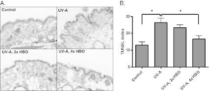
Fig. 4.
TUNEL staining in the skin tissue for DNA fragmentation. a Representative TUNEL-stained images. Magnification, ×200. b Quantification of TUNEL-positive cells per length of epidermis. Cells that showed intense TUNEL staining and an apoptotic morphology (e.g., “rounding-up”) were scored positive (ANOVA with Tukey’s multiple comparison test: *p < 0.01)

