Fig. 7.
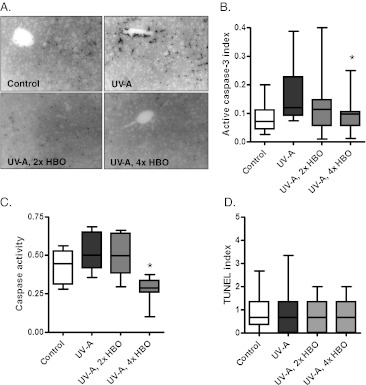
Response of the hepatic tissue to experimental treatment. a Representative cleaved caspase-3 immunohistochemical staining in liver from the different groups. b Quantification of cleaved caspase-3 staining. The ratio of caspase-3-positive cells to the total cells was obtained. The staining index is reduced significantly in the group treated four times per week with HBO (ANOVA and Tukey’s post hoc test: *p < 0.05) c Cleaved caspase-3 activity in cytosolic liver extracts. Enzymatic activity is significantly reduced in animals treated four times per week with HBO relative to the group treated with UV-A alone (Bartlett’s test for equal variance: *p < 0.001). d Quantification of TUNEL staining in liver tissue. TUNEL-positive cells were counted and controlled for field area. No significant differences were found among the groups (ANOVA)

