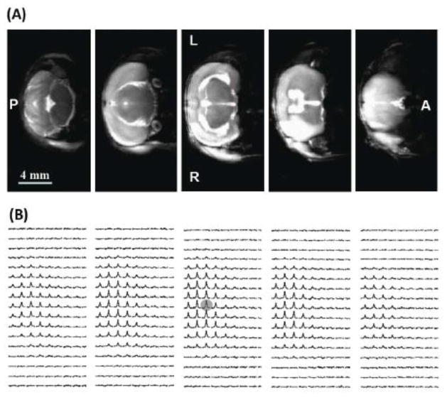
Figure 1.
(A) Multi-slice T2-weighted 1H MRI (8 minutes of total acquisition time and 0.01 μl pixel size), and (B) the corresponding 3D-CSI images of the natural abundance H217O (11 seconds of total acquisition time and 15 μl nominal voxel size) from a representative MCAO mouse brain. The lesions with hyper-intensity in the right brain hemisphere caused by the right MCA occlusion are evident from the anatomic 1H images. L: left side; R: right side; A: anterior image slice; and P: posterior image slice.

