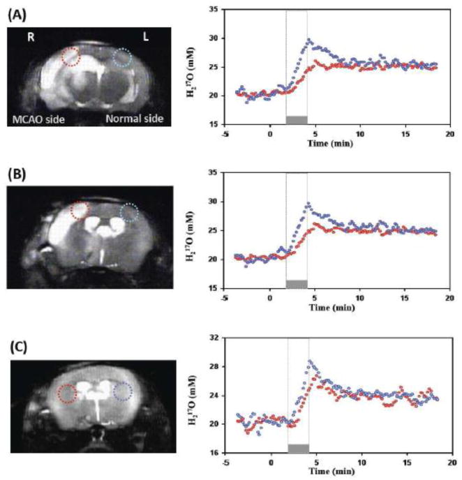
Figure 4.
Comparison of CMRO2 and CBF measurements between the 17O CSI voxels located in the MAC occluded right hemisphere (red circles) and the voxels located in the contralateral left hemisphere of the same mouse brain (blue circles). (A) and (B) show the anatomic images, selected voxels and their corresponding dynamic brain H217O signal changes before, during and after a 2.5-minute 17O2 inhalation from two representative image slices in the same MCAO mouse. Both the slope of 17O signal increase during the inhalation and the exponential decay rate during the post-inhalation phase were substantially smaller in the MCAO affected voxels compared to that corresponding voxels in intact hemisphere, indicating large reductions in both CMRO2 and CBF. (C) Shows the similar results from a different MCAO mouse brain.

