Fig. 3.
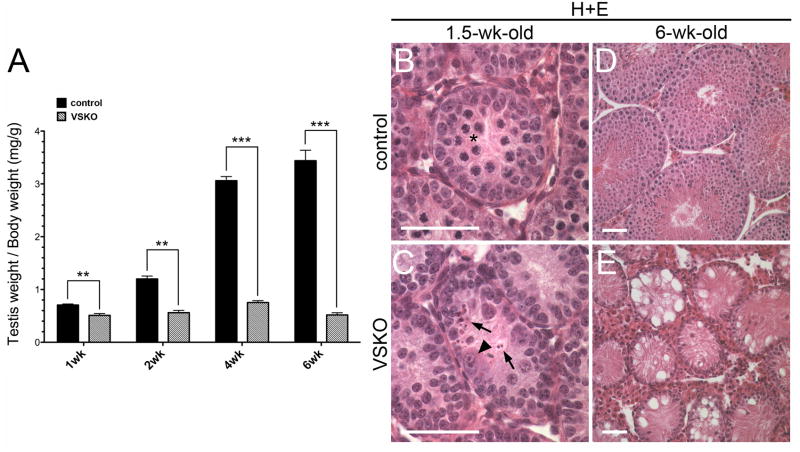
Testes of Vasa-cre; Sin3aΔ/fl male mice are completely depleted of germ cells between 1 and 6 wks of age. A. VSKO testis weight/body weight ratios undergo a significant, progressive reduction from 27.7% to 84.9% between 1 and 6 wks of age, relative to littermate controls (N=3 for each time point; **p<0.01, ***p<0.001). B and C. Seminiferous tubule cross-sections of 1.5-wk-old testes stained with H+E; asterisk denotes meiotic spermatocytes in control section (B). Arrowhead identifies enlarged, abnormal germ cell and arrows denote cell remnants in VSKO section (C). D and E. Cross-sections of 6-wk-old seminiferous tubules stained with H+E; while control sections contain germ cells at all stages and in expected numbers (D), VSKO sections are completely devoid of germ cells, exhibiting a ‘Sertoli cell only’ phenotype (E). Scale bars = 50 μm.

