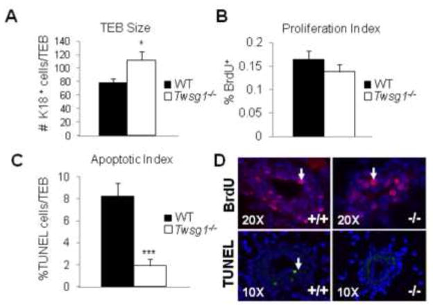
Fig. 7. Twsg1−/− TEBs show an increase in luminal epithelial (K18) cell number and a decrease in apoptosis.
(A) K18 positive cells were counted within TEBs and compared between WT and Twsg1−/−. A significant increase in cell number was observed in Twsg1−/− compared to WT. (B) BrdU positive cells were counted within the same compartment and no significant change in proliferation was observed. (C) TUNEL staining revealed a significant decrease in the number of apoptotic cells within the TEB of Twsg1−/− MGs. (D) Representative images for BrdU (pink), K18 (green) and TUNEL (green) staining. * p<0.05, *** p<0.001

