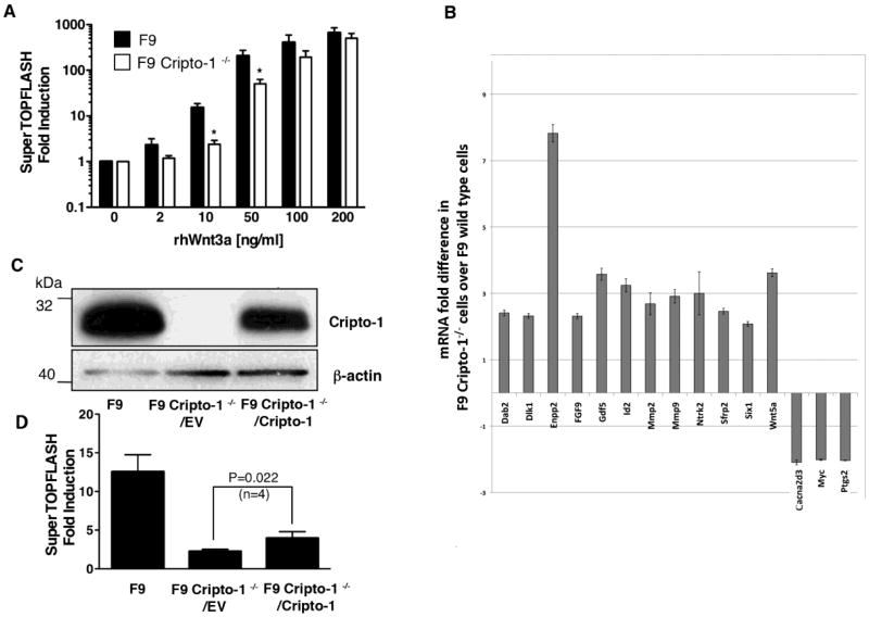
Fig. 8.
Wnt/ β-catenin signaling in F9 and F9 Cripto-1-/- cells. (A) Luciferase reporter assay in F9 and F9 Cripto-1-/- cells transiently transfected with Tcf/Lef responsive SuperTOPFLASH luciferase reporter vector. After transfection, cells were stimulated with different concentrations of rhWnt3a (B) Wnt signaling molecules real time PCR array in F9 and F9 Cripto-1-/- cells. Cells were treated with/without rhWnt3a (100 ng/ml) for 18 h, cDNA was synthesized from total RNA and real time PCR array was performed according to manufacturer's instructions. (C) and (D) Re-expression of Cripto-1 in F9 Cripto-1-/- cells partially rescues sensitivity of F9 Cripto-1-/- cells to rhWnt3a stimulation. (C) A lentiviral Cripto-1 expression vector or a control empty vector were infected into F9 Cripto-1-/- cells, stably expressing lentiviral vectors cells were established and Cripto-1 expression was assessed by Western blot analysis. (D) F9 Cripto-1-/- cells, stably expressing a lentiviral Cripto-1 expression vector or a control empty vector, were compared to F9 control cells in a SuperTOPFLASH luciferase assay after rhWnt3a stimulation (10 ng/ml). P value was calculated using Student's t test.

