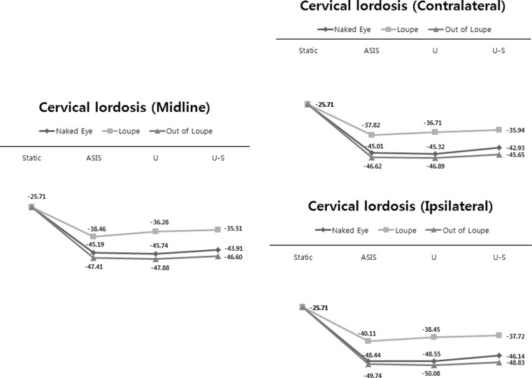
Fig. 5.
Cervical lordosis (αC) measured during the three different visualize methods (naked eye, loupe, out of loupe) and during three different operating table heights (ASIS anterior superior iliac spine, U umbilicus, U–S midpoint of umbilicus and sternum) under three different views (midline, ipsilateral, and contralateral)

